
FIGURE 1.
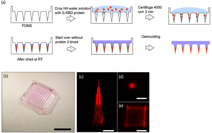
Fabrication and characterization of hyaluronic acid (HA) microneedles (MNs): (a) Illustration of MN fabrication. (b) Images of HA MNs containing Alexa Fluor‐546 rabbit IgG protein (scale bar, 5 mm) (c–e) Confocal images of HA MN tips with Alexa Fluor‐546 rabbit IgG protein at different positions (scale bar c, 200 μm, scale bar d–f, 100 μm)