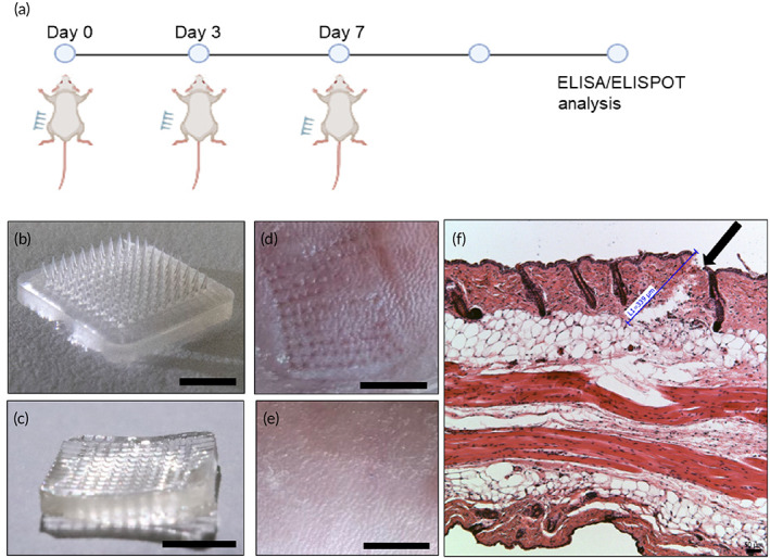
FIGURE 2.

Vaccination with S‐receptor‐binding domain (RBD) microneedles (MNs): (a) Illustration of vaccination process. Image of MN‐RBD patch (b) before injection (tips present) (scale bar, 5 mm) and (c) after injection (tips absent) (scale bar, 5 mm). Image of penetrated mouse skin (d) immediately after injection (scale bar, 5 mm) and (e) 24 h after MN injection (scale bar, 5 mm). (f) Histological image of paraffin‐embedded sections of MN‐penetrated mouse skin. Black arrow indicates site of skin penetration by MNs (scale bar, 50 μm)
