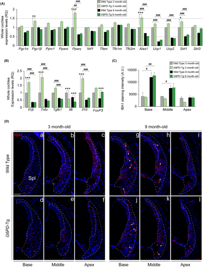
Figure 4.

Inflammatory response and macrophage infiltration in the cochlea. (a and b) RT‐qPCR gene expression levels of mitochondrial biogenesis program genes and pro‐ and anti‐inflammatory cytokines from 3 cochleae pools per condition of 3‐ and 9‐month‐old mice, respectively. Expression levels were calculated as 2−ΔΔCt (RQ), using 18s as a reference gene and normalized to WT data. Data presented as mean ± SEM of triplicate samples. (C) IBA1 staining intensity of the spiral ligament (n = 3 WT, n = 3 G6PD‐Tg). Values are presented as mean ± SEM. Statistical significance was analyzed by Student's t test: *G6PD‐Tg vs WT, # 9‐month‐old mice vs 3‐month‐old mice (*, #p < 0.05; **, ##p < 0.01; ***, ###p < 0.001). (D) Representative cochlear cross‐cryosections immunolabeled for IBA1 showing the spiral ligament (Spl) of the basal, middle, and apical turns (outlined) of both genotypes of 3 (a–f) and 9‐month‐old (g–l) mice. Arrowheads highlight positive staining of macrophages. Scale bar: 25 µm
