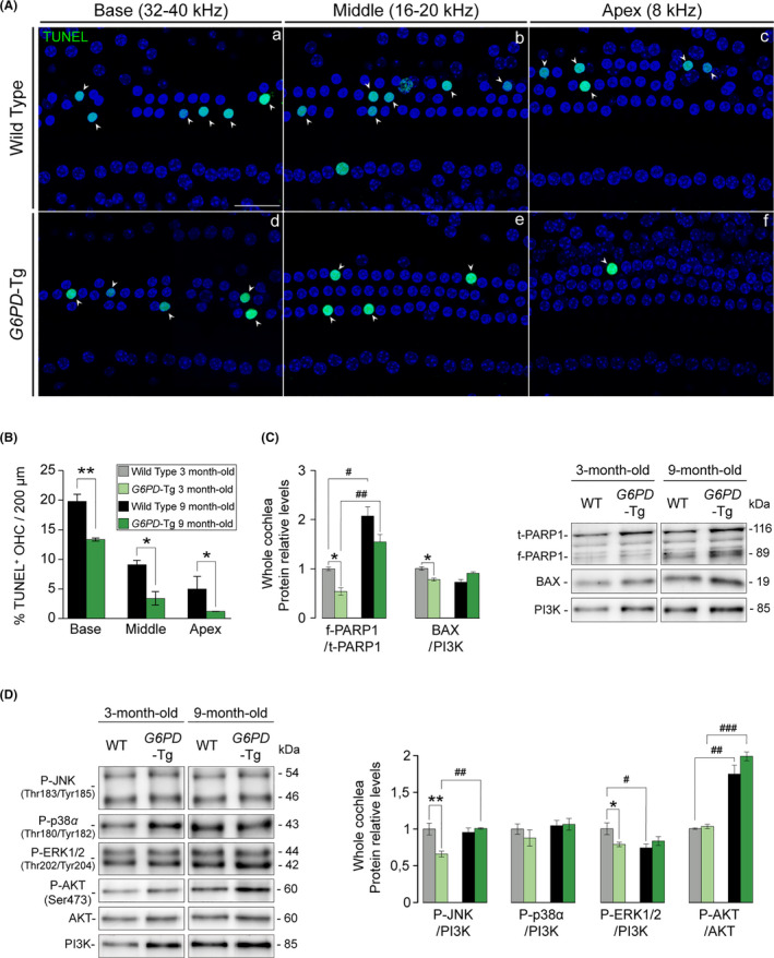
Figure 5.

Diminished apoptotic cell death in 9‐month‐old G6PD‐Tg mice. (A) TUNEL assay of organ of Corti whole mounts of 9‐month‐old mice. Representative confocal images of basal (32–40 kHz), middle (16–20 kHz), and apical (8 kHz) regions of the organ of Corti are shown. Arrowheads indicate TUNEL+ staining. Scale bar: 25 µm. (B) Percentage of TUNEL+ outer hair cells (OHC) in 200‐µm sections of basal (32–40 kHz), middle (16–20 kHz), and apical (8 kHz) regions of the organ of Corti (n = 3 per condition). Statistical significance between genotypes was analyzed by Student's t test (*p < 0.05; **p < 0.01; ***p < 0.001). (c and d) Cochlear protein levels were analyzed by Western blotting of pooled samples from three 3‐ and 9‐month‐old mice per condition. Representative blots and quantifications are shown for pro‐apoptotic and pro‐survival proteins. Protein levels were calculated as a ratio f‐PARP1/t‐PARP1, P‐AKT/AKT, or using PI3K as loading controls, and then normalized to data from 3‐month‐old WT mice. Values presented as mean ± SEM. Statistical significance between genotypes was analyzed Student's t test: *G6PD‐Tg vs WT, # 9‐month‐old mice vs 3‐month‐old mice (*, #p < 0.05; **, ##p < 0.01; ***, ###p < 0.001)
