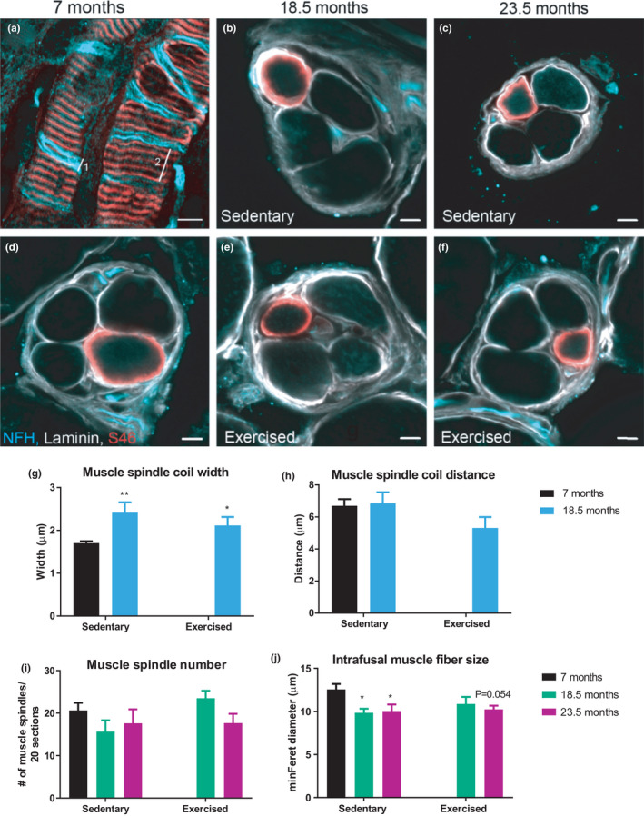
FIGURE 2.

Age affects muscle spindle morphology. (a) Representative image of the longitudinal view of the muscle spindle receptor from EDL whole‐mount, and (b–f) cross‐sectional view from GS muscle. (g, h) Quantification of muscle spindle coil width (line 1 in panel 2a) (g) and coil distance (line 2 in panel 2a) (h) from whole‐mounted EDL muscle. n = 16 muscle spindles from a minimum five animals. (i) Number of muscle spindles per 20 serial sections, and H, the minimal Feret diameter of S46+ intrafusal muscle fibers. n = 2–3 muscle spindles per animal from a minimum five animals. Scale bar = 5 µm. Data show mean ± SEM. For panels (g) and (h), *p < 0.05; **p < 0.01 indicate statistically significant differences between 7 months controls and aged groups (unpaired, two‐tailed Student's t‐test). In panels (i) and (j), *p < 0.05 and p‐values indicates statistically significant differences and statistical trends between 7 months controls and aged groups (age effect) using a two‐way ANOVA comparison
