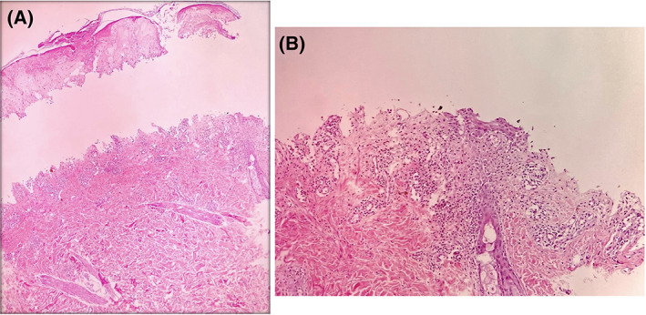
FIGURE 2.

A, Histopathologic examination showed a subepidermal blister with full thickness epidermal necrosis and mild perivascular dermatitis (H&E, ×40); B, Higher magnification shows mild superficial perivascular dermatitis and focal lichenoid tissue reaction (H&E, ×100)
