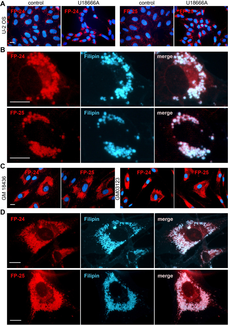
Figure 6.
FP-24 and FP-25 fluorescence in cells with abnormal content of cholesterol. (A) Cholesterol transport in U-2 OS was inhibited by inhibitor U18666A (1 μg/mL) for 48 h, then cells were labelled with indicated probes (100–200 nM) for an additional 24 h and examined. Control cells were treated with vehicle only. (B) Co-localization of FP-24 and FP-25 staining with filipin. U-2 OS cells were treated with inhibitor, labelled with probes, fixed and stained with filipin (50 μg/mL). About 70% of cells displayed significant co-localization of filipin with probe FP-24 (evaluated 107 cells) and 60% with probe FP-25 (evaluated 100 cells). (C) Human fibroblasts carrying mutations in NPC1 cholesterol transporter (clones GM03123E, GM18436) were labelled with probes for 20 h and examined. (D) Co-localization of FP-5 and filipin staining in mutant cell clone GM18436. About 90% of cells displayed significant co-localization of filipin with probe FP-24 (evaluated 70 cells) and 82% with probe FP-25 (evaluated 60 cells). Scale bars 10 μm.

