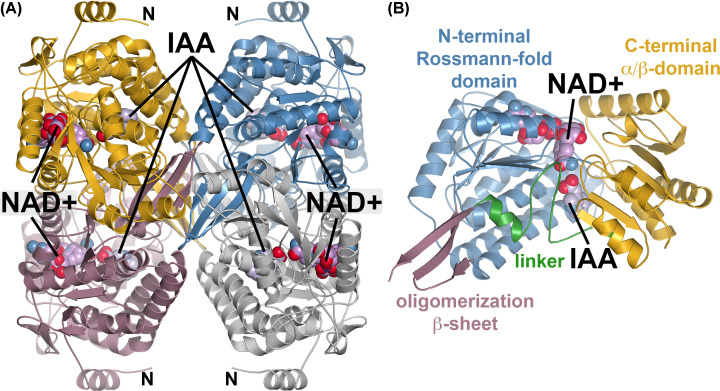
Figure 2. Overall three-dimensional structure of the AldA(C302A)•NAD+•IAA complex.
(A) Tetrameric assembly of the AldA C302 mutant. Each monomer is colored individually with the locations of bound NAD+ and IAA indicated. The N-terminus of each monomer is also noted. (B) Domain organization of an AldA monomer. The view is slightly rotated from that in (A). The N-terminal Rossmann-fold (blue), C-terminal α/β domain (gold), oligomerization β-sheet (rose), and domain linker (green) are indicated and labeled with ligand positions indicated.

