Fig. 5.
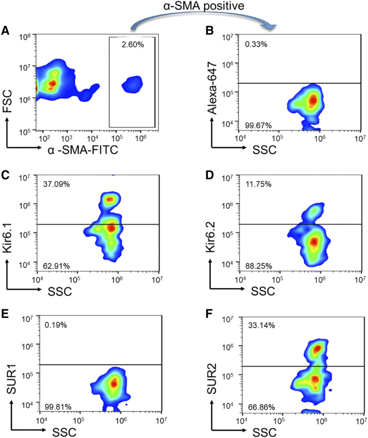
Expression of KATP channel subunits in LMCs. (A) Mesenteric LVs were digested and coincubated with a smooth muscle protein marker, α-SMA–FITC. A gate was set around the cells staining positive for α-SMA. This gate is used in all subsequent panels. (B) Cells selected from the α-SMA positive gate were coincubated with Alexa-647 conjugated secondary antibody only as a negative control. (C) α-SMA positive-gated cells show immunoreactivity corresponding to Kir6.1. (D) α-SMA positive-gated cells also exhibited Kir6.2 immunoreactivity. (E) Immunoreactivity corresponding to SUR1 protein was not detected in α-SMA positive-gated cells. (F) α-SMA positive-gated cells display immunoreactivity for SUR2. FSC, Forward Scatter; SSC, Side Scatter. Images in this figure are representative of n = 3 determinations.

