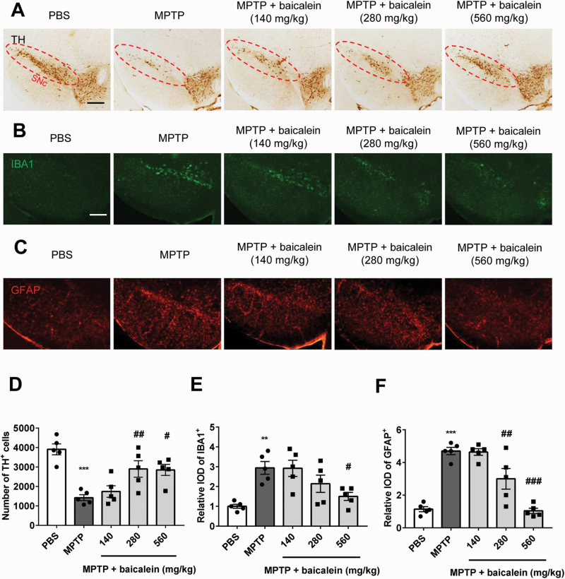
Figure 2.
The protective effect of baicalein on dopaminergic neurons and glial cells. (A) Immunohistochemical staining of tyrosine hydroxylase (TH) neurons in brain substantia nigra compact (SNc) from indicated mice. Scale bar = 100 μm. (B) Immunofluorescence analysis of microglia (ionized calcium binding adapter molecule 1 [IBA1], green) and astrocytes (glial fibrillary acidic protein (GFAP), red) in brain SNc from indicated mice. Scale bar = 100 μm. (C) Quantified cell numbers of TH+ neurons and relative IOD values of IBA1 and GFAP. **P < .01, ***P < .001 vs control group. #P < .05, ##P < .01, ###P < .001 vs MPTP group. Data are expressed as means ± SEM (n = 5/group).

