Figure 5.
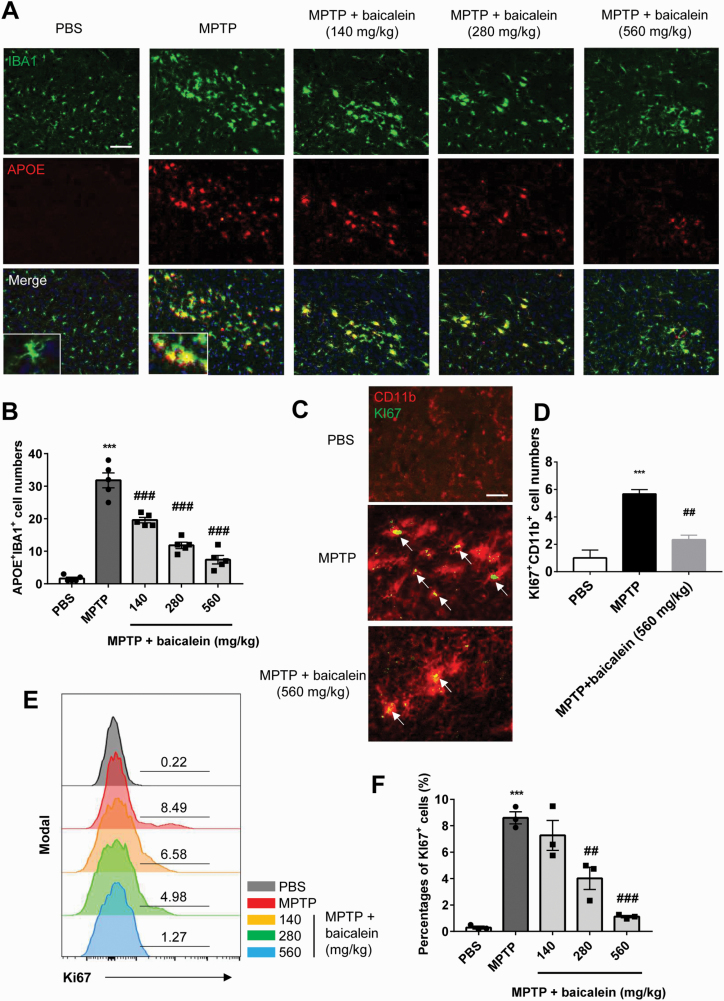
The inhibitory effect of baicalein on proinflammatory microglia. (A–B) Immunofluorescence analysis of ionized calcium binding adapter molecule 1 (green) and APOE (red) cells in brain substantia nigra compact (SNc) from indicated mice. Data are shown as representative images (A) and quantified cell numbers (B) (n = 5/group). Scale bar, 50 μm. (C-D) Immunofluorescence staining of CD11b+ (red) and Ki67+ (green) cells in brain SNc from indicated mice. Data are shown as representative images (C) and quantified cell numbers (D) (n = 3/group). Scale bar = 20 μm. (E–F) Flow-cytometric analysis of Ki67+ cells gated from CD11b+ cells in brain tissue. Data are shown as representative plots (E) and quantified percentages (F) (n = 3/group). ***P < .001 vs control group. ##P < .01, ###P < .001 vs N-methyl-4-phenyl-1,2,3,6-tetrahydropyridine group. Data are expressed as means ± SEM.

