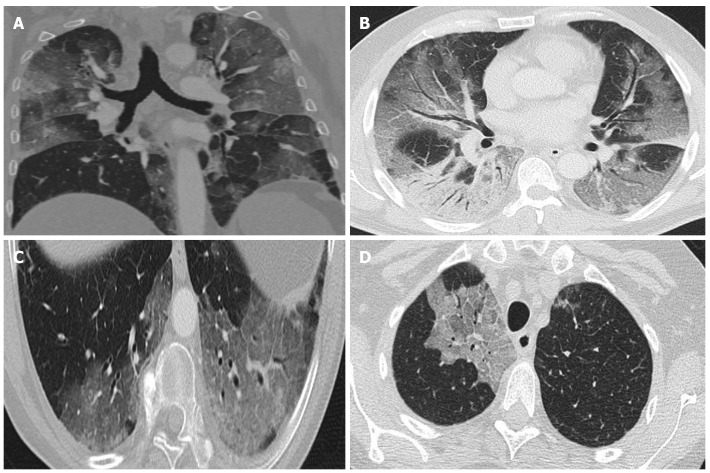
Figure 3.
Computed tomography findings. A: Extensive bilateral ground-glass opacities, one with poorly-defined margins and another with clearly defined borders; B: Multilobar ground-glass opacities and consolidation with air bronchogram in the right lower lobe. Band of subpleural parenchyma respected in the left lung; C: Microvascular dilation sign in the middle of ground-glass opacity in the left lower lobe; D: Crazy-paving pattern in the right upper lobe.

