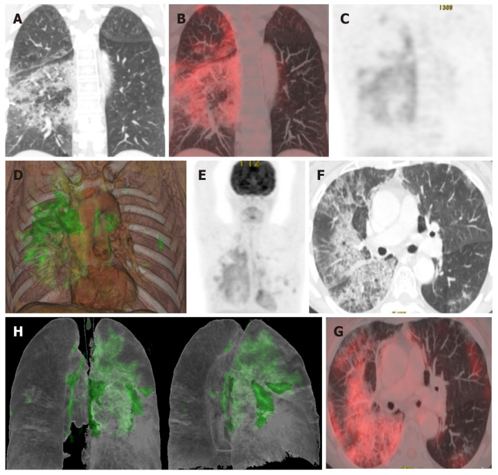
Figure 5.
A 65-year-old patient with a history of invasive lepidic-predominant adenocarcinoma (stage pT1bNxM0) treated with surgery, chemotherapy and radiotherapy. A: Coronal computed tomography showing the crazy-paving pattern (with interstitial septal thickening and increased density of ground-glass opacities) with a markedly asymmetric bilateral distribution, mainly affecting the right side; B: Positron emission tomography-computed tomography (PET-CT) coronal section; C: Metabolic PET; D: Volume rendering 3D PET-CT; E: MIP, PET; B-E: Reveals increased cellular activity [standard uptake value (SUV) 4-6] related to the associated inflammatory process. PET-CT pattern of bilateral coronavirus disease 2019 (COVID-19) viral pneumonitis, predominantly right-sided; F: Axial computed tomography showing crazy-paving pattern (with interstitial septal thickening and increased ground-glass density) with a bilateral, but markedly asymmetric distribution, predominant right-sided; G and H: Axial section and 3D volume rendering from PET-CT metabolic imaging that reveals increased cellular activity (SUV 4-6) related to the associated inflammatory process. PET-CT pattern of bilateral, predominantly right-sided, COVID-19 viral pneumonitis.

