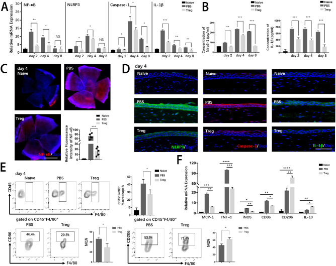
Figure 3.
Treg treatment suppressed excessive inflammation by inhibiting the activation inflammatory pathway and promoting macrophage polarization. (A) The mRNA levels of pro-inflammatory cytokines (NF-κB, NLRP3, caspase-1, and IL-1β) were evaluated by PCR. (B) Protein levels of NLRP3 and IL-1β were analyzed by ELISA. (C) Immunostaining of NF-κB was conducted in whole corneal tissue. Nuclei were counterstained with DAPI. Scar bar: 1 mm. The relative fluorescence intensity of NF-κB analyzed by ImageJ is shown. Each group consisted of five mice. (D) Representative merged immunofluorescence staining images of inflammatory markers (NLRP3, caspase-1, and IL-1β) are presented. Nuclei were counterstained with DAPI. Scar bar: 50 µm. (E) Representative flow cytometric contour plots exhibited infiltration of corneal CD45+ F4/80+ macrophages in naïve and alkali-injured mice on day 4. Representative flow cytometric contour plots showed frequencies of CD45+F4/80+CD86+ (M1) macrophages and CD45+F4/80+CD206+ (M2) macrophages.

