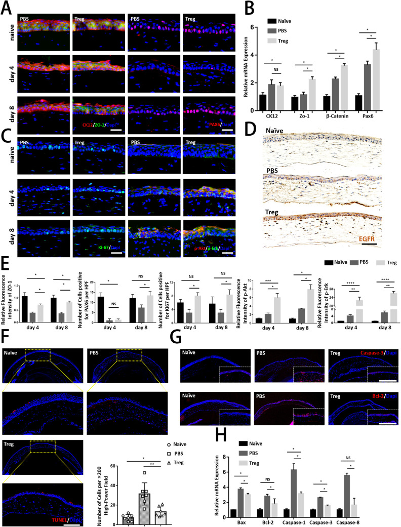
Figure 4.
Tregs recovered cornea structurally and functionally by promoting proliferation and inhibiting the apoptosis of corneal epithelial cells. (A) Representative immunofluorescence micrographs of corneas stained with CK12 (a corneal epithelial marker), ZO-1 (an epithelial tight junction marker), and PAX6 on days 4 and 8 after the injury. Scar bar: 25 µm. (B) Relative mRNA expression of corneal structural and functional markers (CK12, ZO-1, β-Catenin, and Pax6) is shown. (C) The regenerating corneal epithelium was stained with Ki-67 (a proliferation marker), p-Akt, and p-Erk on days 4 and 8 after the injury. Scar bar: 25 µm. (D) Representative immunofluorescence micrographs of corneas stained with EGFR are shown. Scar bar: 50 µm. (E) The panels analyzed the relative fluorescence intensity and number of cells per high-power field (HPF) in above immunofluorescence graphs. (F) TUNEL staining was conducted in corneas. Staining of seven corneas in each group was quantified by calculating the percentage of TUNEL+ cells in each image. Scar bar: 50 µm. (G) Representative immunofluorescence micrographs of corneas stained with caspase-3 and Bcl-2 are shown. Scar bar: 50 µm. (H) Relative mRNA expression of apoptosis genes (Bax, Bcl-2, caspase-1, caspase-3, and caspase-8) is shown.

