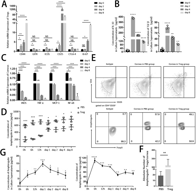
Figure 5.
Tregs had higher levels of functional markers, including amphiregulin after being stimulated by alkali-injured corneas. Freshly isolated Tregs were co-cultured with alkali-burned corneas using the Transwell System. (A) Expression of markers related to Tregs (CD4+CD25+Foxp3+) function and activation (CD44, GITR, ICOS, CD25, CTLA-4, and Ki67) after co-culturing with damaged corneas for 2, 4, and 8 days in vitro was evaluated. (B) The protein levels of anti-inflammatory cytokines IL-10 and TGF-β in culture supernatants were analyzed by ELISA. (C) Relative mRNA expression of pro-inflammatory cytokines in corneas in vitro cocultured with Tregs was examined. (D) Representative flow cytometry plots showing the amphiregulin expression in corneas from PBS and Treg groups (gated on live cells). (E) The lower panel analyzed the number of Areg+ Tregs per cornea. (F) Kinetics protein levels of amphiregulin in alkali-injured corneas at different time points after Tregs or PBS (control) treatment. (G) Concentration of amphiregulin protein in supernatants and in Tregs was examined by ELISA.

