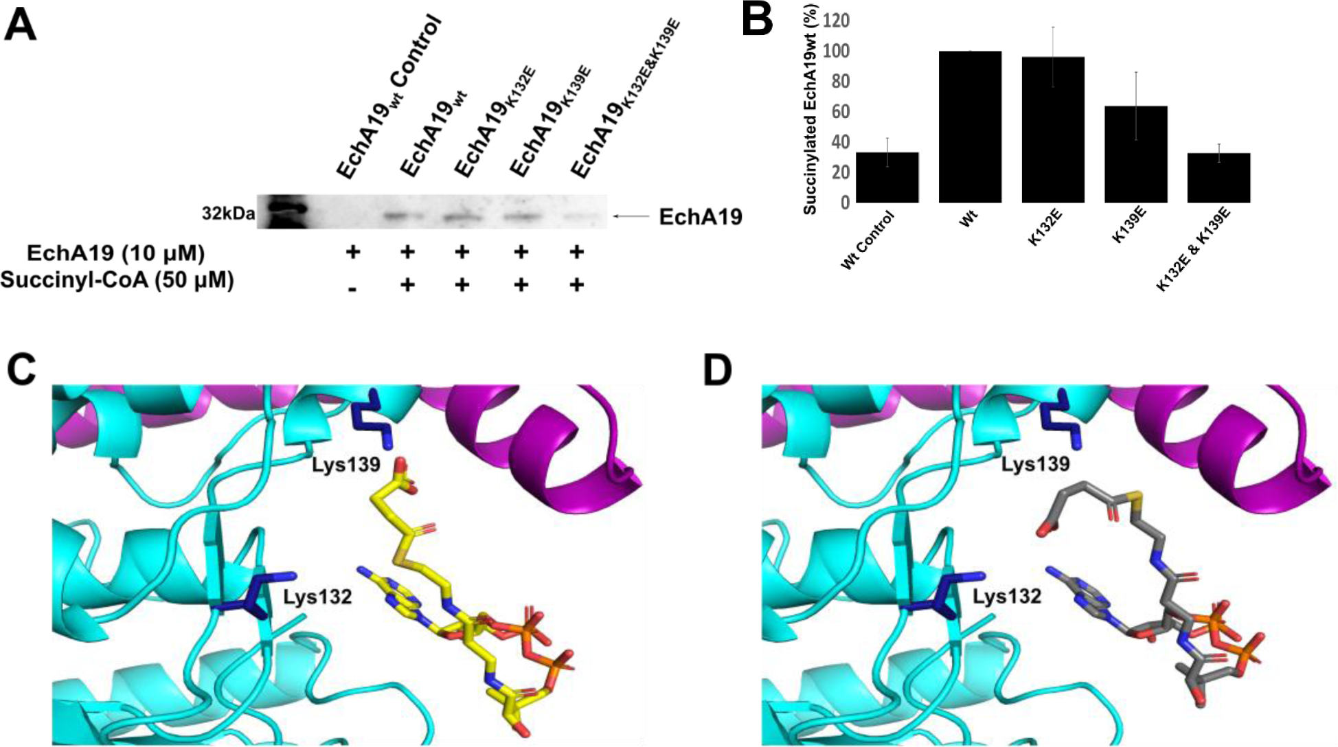
Figure 2. EchA19 enzymes are succinylated at sites near the substrate binding pockets.

A) Recombinant EchA19wt and three lysine mutants (EchA19K132E, EchA19K139E, and EchA19K132E&K139E) were incubated with 50 μM succinyl-CoA at pH 8.1 for 60 min, and the extent of succinylation was monitored with a pan anti-succinyllysine antibody. B) Densitometry analysis of blots from three replicates of experiment shown in (A). Intensity was scaled to 100% for succinylated EchA19wt. Errors shown are the standard error of measurement. C) and D) The active site of EchA19 with succinyl-CoA modeled in the CoA pocket identified in the 3-OCDO-CoA-bound model (Figure 1). Lys132 and Lys139 (dark blue) are located near the CoA binding pocket of EchA19.
