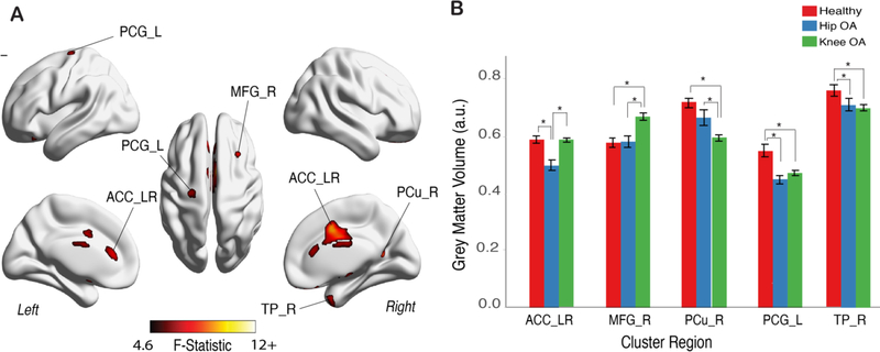
Figure 2. Primary motor cortex, contralateral to OA pain, showed lower volume in both HOA and KOA patients.
A. Grey matter morphological changes assessed by voxel-based morphometry (VBM) after aligning the brain in relation to OA pain (brain x axis left-to-right flipping for left sided KOA and HOA; which renders the left hemisphere to be contralateral to OA in all patients). The three groups were contrasted together, using a 1-way between subjects’ ANCOVA, controlling for age, gender, total ICV and brain flipping. Shown is the F-test statistic map clustering determined using a TFCE uncorrected p-value binarized mask at value of 0.001 and cluster size >66 voxels. Multiple brain regions showed changes in regional volume B. Post-hoc results for GM volume differences for significant clusters, adjusted for age, gender and TICV (Tukey HSD tests at p<0.001); Bars represent the mean grey matter volume and error bars represent the standard error. Left primary motor cortex (precentral gyrus; PCG_L) and right temporal pole (TP_R) presented lower GM volume in both OA groups. Bilateral anterior cingulate cortex (ACC_LR) showed lower GM volume only in HOA, when contrasted with the other two groups. In the KOA group, GM volume was greater for middle frontal gyrus cluster on the right (MFG_R) and lower for right precuneus cortex (PC_R). Further information on thresholded clusters can be found in Table 2. *p<0.001.

