. 2020 Dec 7;9:e61405. doi: 10.7554/eLife.61405

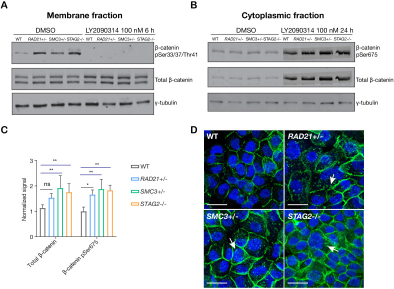
Figure 5. LY2090314-mediated WNT stimulation leads to β-catenin stabilization in cohesin-deficient MCF10A cells.

(A) Immunoblot of the membrane fraction of parental (WT) and cohesin-deficient MCF10A cells shows increased basal level of β-catenin phosphorylation at Ser33/37/Thr41. (B) Immunoblot of the cytoplasmic fraction shows increased level of both total and phosphorylated β-catenin at Ser675 after parental (WT) and cohesin-deficient MCF10A cells were treated with LY2090314 at 100 nM for 24 hr. (C) Quantification of protein levels for total and phosphorylated β-catenin at Ser675. n = 3 independent experiments, mean ± s.d., one-way ANOVA: *p≤0.05, **p≤0.01. (D) Immunofluorescence images show cytosolic accumulation of active β-catenin in cohesin-deficient MCF10A cells treated with LY2090314 100 nM for 24 hr, relative to parental (WT) MCF10A cells. White arrows indicate puncta of β-catenin (pSer675). Scale bar = 25 μM. Full length blots and molecular size markers are available for A,B in Figure 5—source data 1. Source quantification data is available for C in Figure 5—source data 2.

Figure 5—source data 1. Untrimmed blots for Figure 5A, B.
Figure 5—source data 2. Quantitation of blots in Figure 5A, B.
Figure 5—source data 3. Untrimmed blots for Figure 5A, B.
Figure 5—source data 4. Untrimmed blots for Figure 5A, B.

Figure 5.

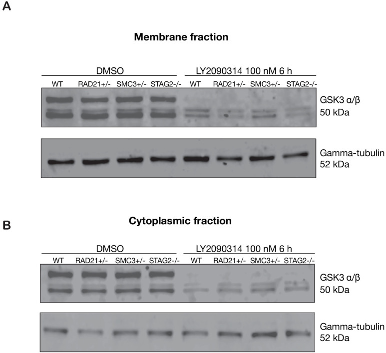
Figure 5—figure supplement 1. GSK3 levels are unaffected in cohesin-deficient MCF10A cells.

Figure 5—figure supplement 1.

Anti-GSK3 immunoblot of membrane fraction from parental MCF10A (WT) or cohesin-deficient cells treated with either DMSO or 100 nM LY2090314. (A) Membrane fraction after 6 hr of treatment with DMSO, or with LY2090314. (B) Cytoplasmic fraction after 6 hr of treatment with DMSO, or with LY2090314. Untrimmed blots and molecular size markers are available for A,B in Figure 5—source data 3.
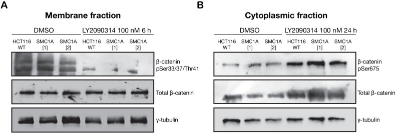
Figure 5—figure supplement 2. LY2090314-mediated WNT stimulation leads to increased β-catenin stabilization in SMC1A mutant HCT116 cells.

Figure 5—figure supplement 2.

Unmodified HCT116 cells (WT), and cells modified to contain SMC1A mutations: SMC1A (1), c.2027A > G; SMC1A (2), c.2479 C > T, were treated with DMSO or LY2090314 at 100 nM for 24 hr. (A) Immunoblot of the membrane fraction of HCT116 wild type (WT) and SMC1A mutant-expressing HCT116 cells. The proteasome-targeted form of β-catenin phosphorylated at Ser33/37/Thr41 is slightly less abundant in the SMC1A mutants, and this phospho isoform is degraded in all cells following treatment with LY2090314. (B) Immunoblot of the cytoplasmic fraction shows increased basal level of phosphorylated β-catenin at Ser675 in the SMC1A mutant-expressing cells compared with HCT116 WT, and LY2090314 treatment markedly increased total β-catenin in the SMC1A mutants compared with HCT116 WT. Untrimmed blots and molecular size markers are available for A,B in Figure 5—source data 4.
Figure 5—figure supplement 3. WNT3A phenocopies LY2090314-mediated β-catenin accumulation in the cytoplasm.
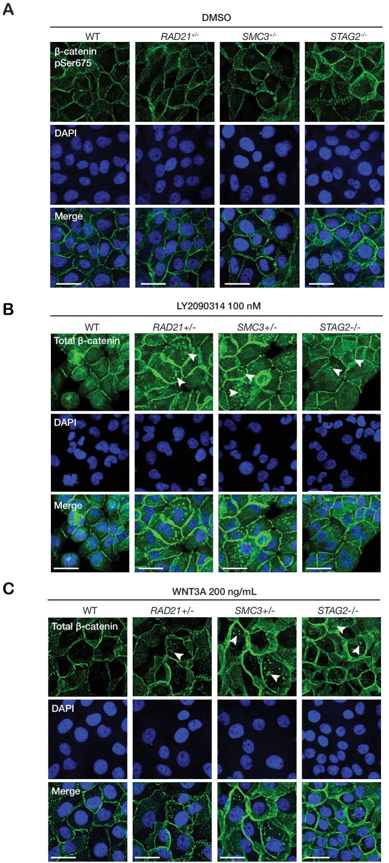
Figure 5—figure supplement 3.

Immunofluorescence images show that (A) parental MCF10A (WT) or cohesin-deficient cells treated with 0.5% DMSO have no β-catenin puncta. In contrast, 24 hr of treatment with both (B) LY2090314 and (C) WNT3A leads to formation of β-catenin puncta in the cytoplasm of cohesin-deficient cells (arrows), but not in parental MCF10A (WT) cells. Scale bar = 25 µm.