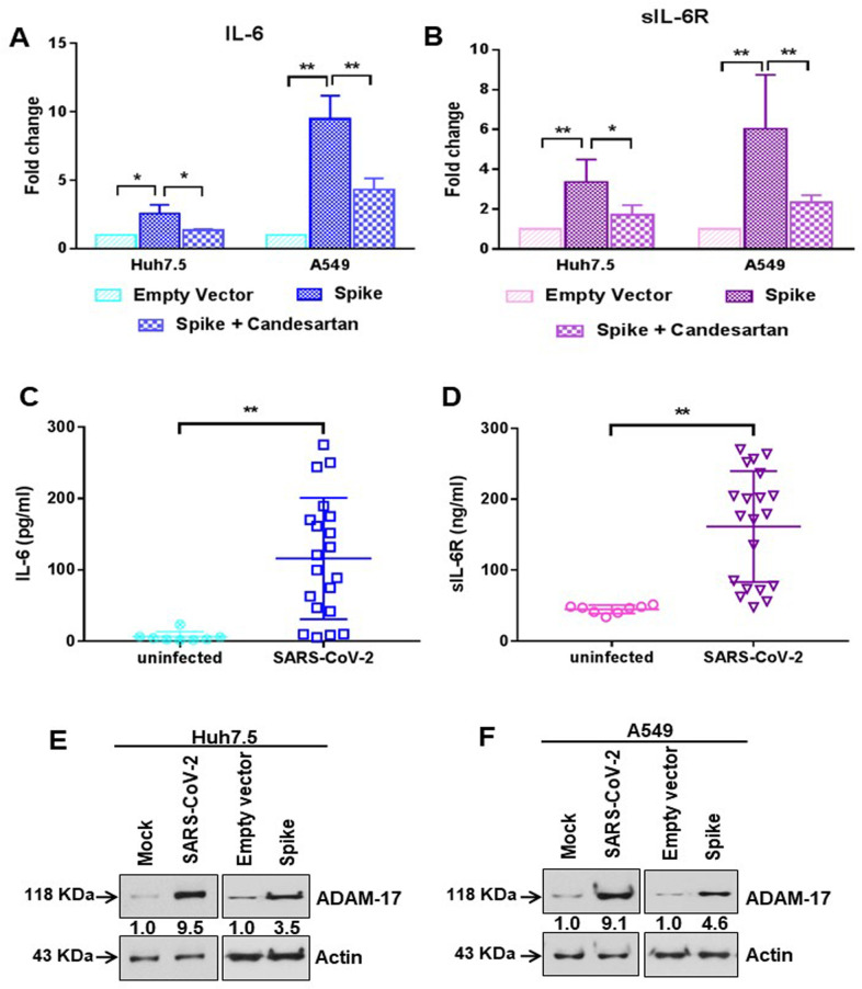
Fig 4. SARS-CoV-2 spike protein stimulates IL-6 and soluble IL-6R production.
(A) The extra-cellular level of IL-6 was measured by ELISA from culture supernatant of Huh7.5 and A549 cells after transfection of SARS-CoV-2 spike gene construct with or without Candesartan cilexetil treatment. (B) The extra-cellular level of soluble IL-6R was similarly measured by ELISA from culture supernatant of Huh7.5 and A549 cells after transfection of SARS-CoV-2 spike gene construct with or without Candesartan cilexetil treatment as shown in panel A. The results are presented as means ± standard deviations. ‘*’ and ‘**’ represent statistical significance p<0.05 and p<0.005, respectively. (C) The level of IL-6 was measured by ELISA from the serum samples of SARS-CoV-2 infected patients (n = 20) and uninfected healthy volunteers (n = 8). (D) The level of soluble IL-6R was similarly measured by ELISA from the serum samples of SARS-CoV-2 infected patients (n = 20) and uninfected healthy volunteers (n = 8) as shown in panel C. The results are presented as means ± standard deviations. ‘*’ and ‘**’ represent statistical significance p<0.05 and p<0.005, respectively. (E, F) Western blot analysis of ADAM-17 expression in Huh7.5 and A549 cell lysates prepared after 48 h of mock-treated or infected with SARS-CoV-2 virus, or transiently transfected with empty vector or SARS-CoV-2 spike gene construct. Expression level of actin in each lane is shown as a total protein loading control for comparison.

