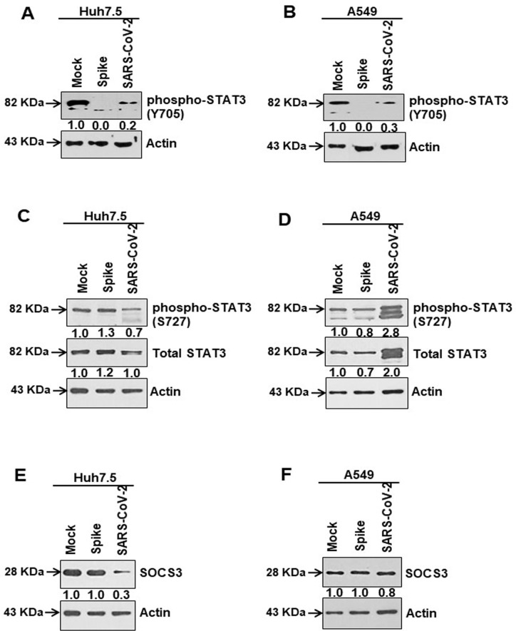
Fig 6. SARS-CoV-2 spike protein causes inhibition of tyrosine phosphorylation of STAT3.
(A, B) Western blot analysis of phospho-STAT3 (Tyr705) and total STAT3 expression in Huh7.5 and A549 cell lysates prepared after 48 h of mock-treated or transient transfection of SARS-CoV-2 spike gene constructs or infection with SARS-CoV-2 virus. (C, D) Western blot analysis of phospho-STAT3 (Ser727) and total STAT3 expression in Huh7.5 and A549 cell lysates prepared after 48 h of mock-treated or transiently transfection of SARS-CoV-2 spike gene construct or infection with SARS-CoV-2 virus. (E, F) Western blot analysis of SOCS3 expression in Huh7.5 and A549 cell lysates prepared after 48 h of mock-treated or transiently transfected with SARS-CoV-2 spike constructs or infected with SARS-CoV-2 virus. Expression level of actin in each lane from the same gel is shown as a total protein loading control for comparison.

