
Fig. 5. Sex differences in Npy mRNA expression and NPY protein densities following oxycodone CPP.
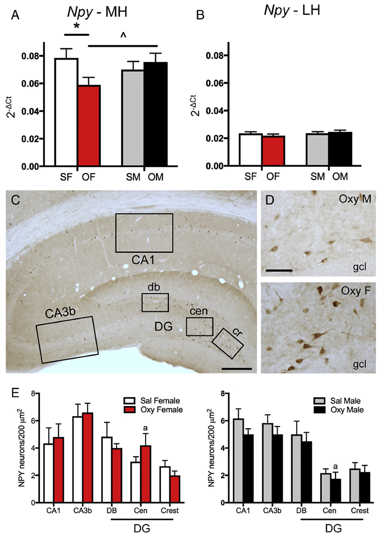
A. In the medial hippocampus (MH), Sal-females (SF) had significantly greater Npy expression than Oxy-females (OF). Moreover, Oxy-females had a trend towards less Npy expression compared to Oxy-males. B. In the lateral hippocampus (LH), there were no differences in Npy expression between any groups. C. Low magnification photomicrograph of the rat dorsal hippocampus shows NPY-labeled cells scattered throughout the hippocampus. Boxed areas show regions sampled for quantitative analysis: CA1, CA3b and the dorsal blade (db), central hilus (cen) and crest (cr) of the dentate gyrus (DG). Bar = 500 μm. D. Representative light microscope micrographs show fewer NPY-labeled neurons in the central hilus of the dentate gyrus of an Oxy-male compared to an Oxy-female. gel, granule cell layer Bar = 50 μm. E. There was a trend for more NPY-labeled cells in the central hilus of Oxy-females compared to Oxy-males. * p < 0.05, ap = 0.055.