
Fig. 8. Sex differences in Mapk1 expression and MAPK and pMAPK protein densities following oxycodone CPP.
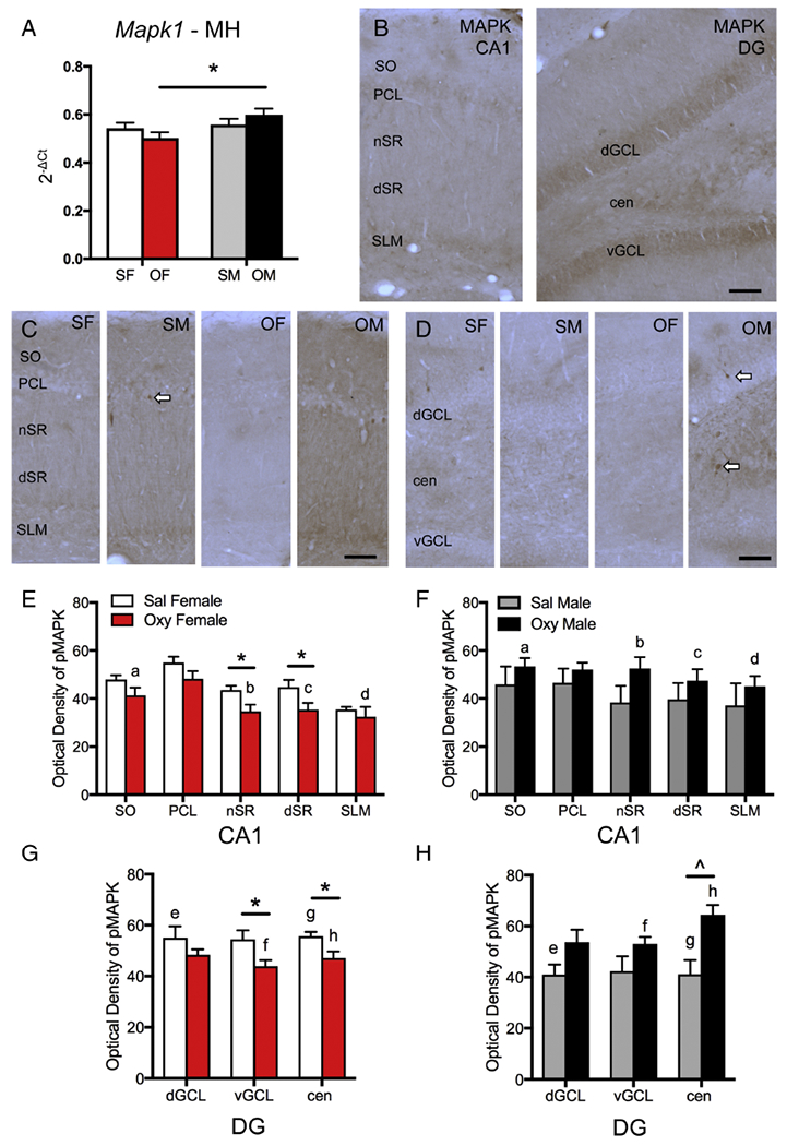
A. In the medial hippocampus (MH), Mapk1 expression is significantly lower in Oxy-females (OF) compared to Oxy-males (OM). B. In CA1 (left), MAPK-ir is densest in the pyramidal cell layer (PCL) and stratum lacunosum-moleculare (SLM) but is also found throughout the other lamina (SO, nSR, dSR). In DG (right), MAPK-ir is dense in both the dorsal and ventral granule cell layers (dGCL, vGCL) and in the central hilus (cen). C. Representative light microscope photographs show distribution of pMAPK-ir in the CA1 region. D. Representative light microscope photographs show distribution of pMAPK-ir in the dentate gyrus. SF, Sal-female; SM, Sal-male. Arrows indicate examples of pMAPK-labeled cells. Bars = 25 μm. E & F. In CA1, the density of pMAPK-ir significantly decreased in the nSR and dSR of Oxy-females compared to Sal-females. Moreover, Oxy-females had significantly lower densities of pMAPK-ir in SO and nSR and a trend towards lower density of pMAK-ir in dSR and SLM compared to Oxy-males. G & H. In the dentate gyrus, Sal-females had significantly higher densities of pMAPK-ir the dGCL and central hilus compared to Sal-males. Sal-females had significantly higher densities of pMAPK-ir than Oxy-females in the vGCL and in the central hilus. Sal-males had a trend towards lower density of pMAPK-ir than Oxy-males in the central hilus. Oxy-females compared to Oxy-males had significantly lower densities of pMAPK-ir in the vGCL and central hilus. *p < 0.05, ap = 0.034, bp = 0.013, cp = 0.061, dp = 0.063, ep = 0.040; fp = 0.030, gp = 0.041; hp = 0.006.