Figure 1.

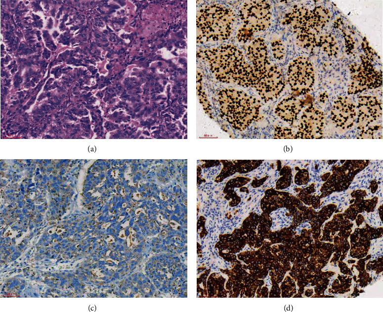
The HE staining and IHC images from lung cancer patients with liver metastasis. (a) Lung cancer (HE ×200). (b) TTF-1(+) (SP ×200). (c) NapsinA(+) (SP ×200). (d) CK7(+) (SP ×200).

The HE staining and IHC images from lung cancer patients with liver metastasis. (a) Lung cancer (HE ×200). (b) TTF-1(+) (SP ×200). (c) NapsinA(+) (SP ×200). (d) CK7(+) (SP ×200).