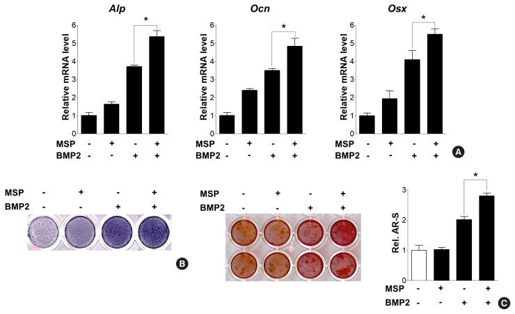
Fig. 3.
Macrophage-stimulating protein (MSP) enhanced bone morphogenetic protein 2 (BMP2)-induced osteoblast differentiation and calcium deposition. (A) Quantitative reverse transcription-polymerase chain reaction (qRT-PCR) of primary osteoblasts were treated with MSP (100 ng/mL) in the presence or absence of BMP2 (50 ng/mL) for 3 days. (B) Alkaline phosphatase (Alp) staining of cells were treated with MSP or/and BMP2 for 5 days, as described above. (C) After 9 days, the cells were stained with alizarin red staining solution. To quantify calcium deposition, the stained samples were treated with 10% cetylpyridinium chloride, and the absorbance of the eluting solution was measured at 540 nm using a spectrophotometer. *P<0.05.

