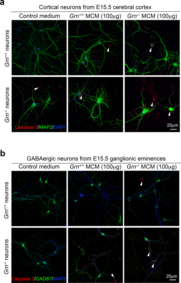
Extended Data Figure 6 |. Grn−/− microglial conditioned media (MCM)-induced cell death in Grn+/+ and Grn−/− cortical neurons and GABAergic neurons.
a. Representative confocal images of Grn+/+ and Grn−/− cortical neurons treated with control media, Grn+/+ MCM or Grn−/− MCM (100μg/ml) overnight. Immunofluorescent stains are performed using antibodies for MAP2 (green) and cleaved caspase 3 (red). Nuclei are highlighted using DAPI. b. Representative confocal microscopic images of GE-derived Grn+/+ and Grn−/− GABAergic interneurons treated with control media, Grn+/+ MCM or Grn−/− MCM (100μg/ml) overnight. Immunofluorescent stains are performed using antibodies for GAD67 (green) and cleaved caspase 3 (red). Nuclei are highlighted using DAPI.

