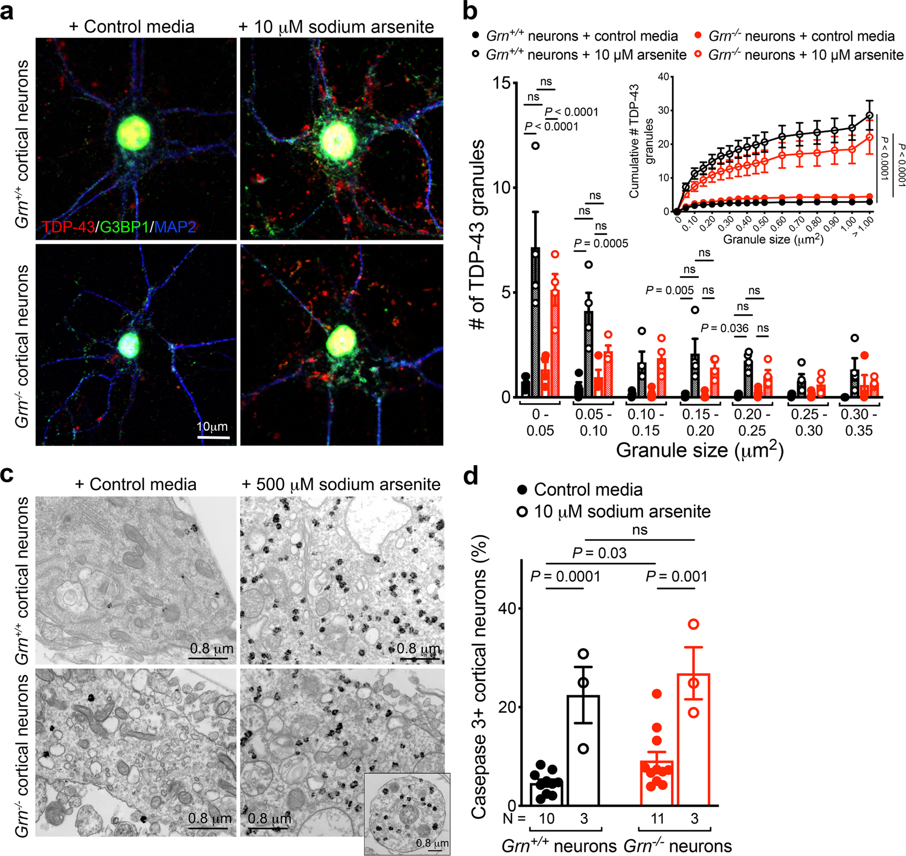
Extended Data Figure 9 |. Sodium arsenite-induced TDP-43 granules in Grn+/+ and Grn−/− cortical neurons do not colocalize with G3BP1+ stress granules.

a. Sodium arsenite treatment (10 μM, 1 hr) induces prominent TDP-43 granules (red) and G3BP1+ granules (green) in Grn+/+ and Grn−/− cortical neurons. However, the TDP-43 granules and G3BP1+ granules show no evidence of colocalization in these neurons. b. Quantification using NIH ImageJ shows that the majority of TDP-43 granules are smaller than 0.05 μm2. In contrast to Grn−/− MCM treatment, sodium arsenite induces similar TDP-43 granule formation in Grn+/+ and Grn−/− cortical neurons. Images in panel a and quantification in panel b were obtained from four independent cultures. Data represent mean ± s.e.m.. Statistics use two-way ANOVA with multiple comparisons. c. Immunogold electron microscopy (IEM) reveals that TDP-43 granules induced by sodium arsenite (500 μM) have morphology similar to those in Grn−/− thalamic neurons (Figure 2d) and Grn−/− cortical neurons treated with Grn−/− MCM (Extended Data Figure 8d). At least 8 IEM images were analyzed from 2 independent cultures per condition. d. Grn+/+ and Grn−/− cortical neurons are equally vulnerable to sodium arsenite treatment (10 μM, 1 hr). Data represent mean ± s.e.m. N indicates the number of independent cultures. Statistics use two-tailed unpaired Student’s t test, ns, not significant.
