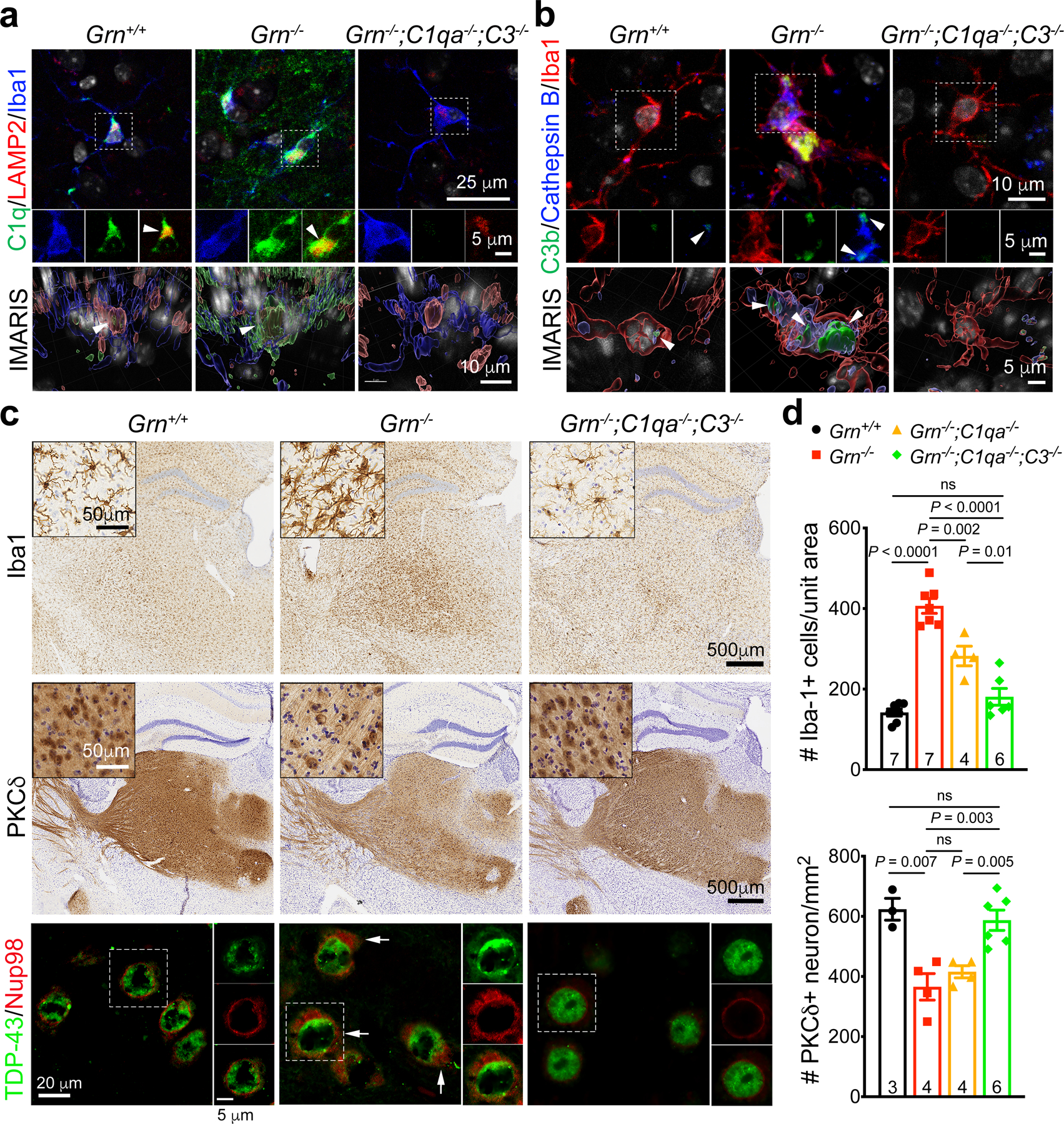
Figure 4 |. Complements C1q and C3b promote TDP-43 granule formation and neurodegeneration in Grn−/− mice.

a–b. Confocal and IMARIS 3D images of C1q and C3b proteins in the cytoplasm of Th-MG in 12 months old Grn+/+, Grn−/− and Grn−/−;C1qa−/−;C3−/− mice. Small amounts of C1q and C3b proteins are detected in Grn+/+ Th-MG where they colocalize with LAMP2+ or Cathepsin B+ vesicles (left panels), whereas much more abundant C1q and C3b proteins are present in Grn−/− Th-MG where these two proteins show overlap with LAMP2+ and Cathepsin B+ vesicles (arrowheads). No C1q or C3b signals are detected in Grn−/−;C1qa−/−;C3−/− Th-MG. Similar results were obtained from n=3 mice per genotype. c. Immunohistochemistry show a near complete rescue of microgliosis (1st row) and PKCδ+ neuron loss (2nd row) in the ventral thalamus of 12 months old Grn−/−;C1qa−/−;C3−/− mice. Insets are higher magnifications for Iba1+ microglia and PKCδ+ neurons. Confocal images show the presence of cytoplasmic TDP-43 and Nup98 in Grn−/− neurons (arrows, 3rd row), but not in Grn−/−;C1qa−/−;C3−/− neurons. d. Stereology quantification of microglia and PKCδ+ neuron in 12 months old Grn+/+, Grn−/−, Grn−/−;C1qa−/− and Grn−/−;C1qa−/−;C3−/− mice. Data represent mean ± s.e.m. The number of mice for each genotype is indicated at the bottom of each graph. Statistics use two-tailed unpaired Student’s t test, ns, not significant.
