
Figure 3.
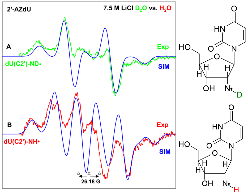
EPR spectrum of aminyl radical formed in matched samples (1 mg/mL of each compound) via radiation-produced (absorbed dose = 500 Gy at 77 K) one-electron addition by γ-irradiation of 2 (A) dU(C2′)-ND• (green color) in 7.5 M LiCl (D2O), (B) dU(C2′)-NH• (red color) in 7.5 M LiCl (H2O) color after visible illumination of each sample by using a photoflood lamp at 77 K for 15 minutes to remove the uracil anion radical by photoejection of the excess electron. Figure 3(A) is the same one shown in Figures 1A and 2A. The simulated spectra (blue) are superimposed on the top of the experimentally recorded spectra for comparison. Each experimentally recorded spectrum shown in Figure 2 was obtained after subtraction of the line components of Cl2•− spectrum following our previous works on the isolation of 77 K RNH• spectrum.7–9,49–51