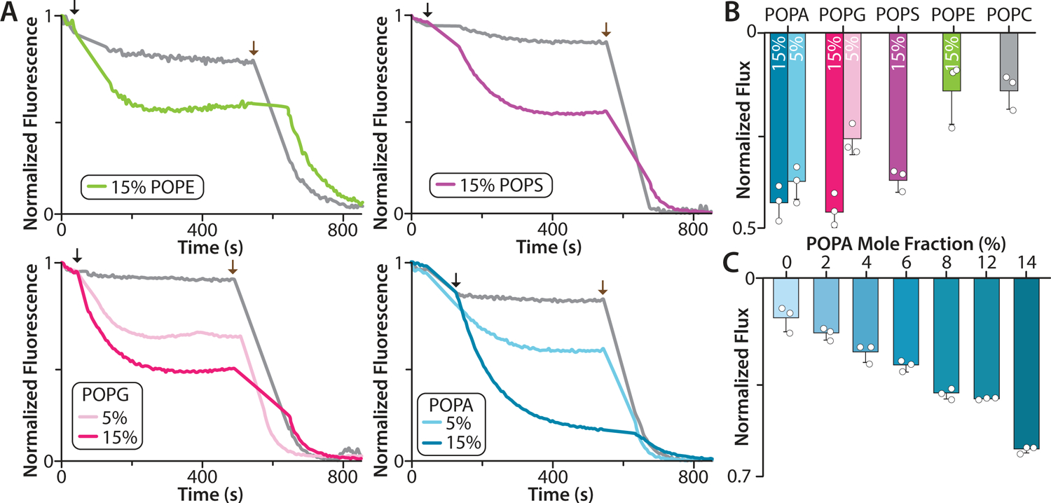
Figure 4. Liposome potassium flux assays of K2P4.1a reconstituted in defined lipid environments.

A) Flux assay traces for K2P4.1a reconstituted in POPC doped with POPG, POPS, POPE, or POPA. Empty liposomes of the same lipid composition are shown in grey. The addition of the proton ionophore carbonyl cyanide m-chlorophenylhydrazone and the potassium ionophore valinomycin is denoted by black and brown arrows, respectively. The mole fraction (%) of doped lipid is shown in the inset. B) Normalized K2P4.1a flux in POPC with 15% POPG, POPS, POPS, or POPA. The normalized flux for POPC with 5% POPG or POPA is shown. C) K2P4.1a potassium efflux in POPC containing different mole fractions of POPA. Data are presented as the mean ± standard deviation (n = 3) for three independent experiments (white dots).
