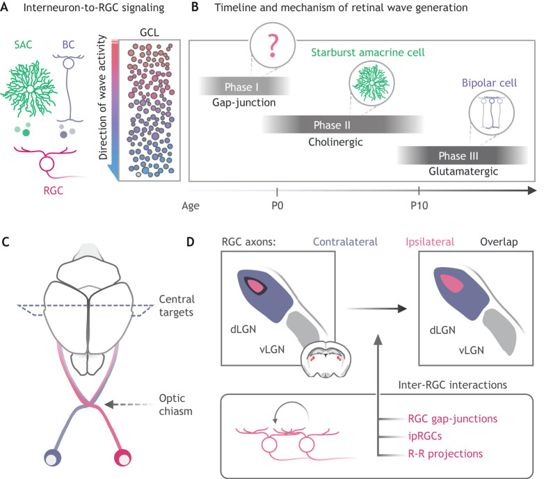
Fig. 3.
Inter-RGC interactions shape early retinal activity. (A) Retinal waves are mediated through release of acetylcholine from starburst amacrine cells (SACs), glutamate via bipolar cells (BC), or through gap-junction coupling between retinal ganglion cells (RGCs). The activity of these cells drives waves of depolarization across the ganglion cell layer (GCL, right) to central targets involved in binocular vision (C,D) and refines synapses between RGCs and their targets. (B) Over developmental time, retinal waves are generated via distinct mechanisms (cell within circle) with partial overlap. Phase I (∼E16-P0), gap-junction-mediated waves; phase II (∼P0-P10), cholinergic waves; and phase III (∼P10-P14), glutamatergic waves. (C) RGCs from each retina extend axons towards the brain and cross at the optic chiasm or continue along the same side (pink versus purple axons). (D) At the visual thalamus (dLGN, dorsal lateral geniculate nucleus; vLGN, ventral lateral geniculate nucleus), retinal waves drive refinement of retinal projections from the eye on the same side (ipsilateral; pink) or opposite side (contralateral; purple). Inter-RGC interactions (bottom) are considered to drive a portion of this refinement process, promoting segregation of projection and thus less overlap (black regions in dorsal LGN). ipRGCs, intrinsically photosensitive RGCs; R-R projections, retino-retinal projections.

