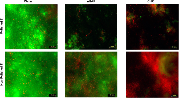
FIGURE 3.
Fluorescence microscopic investigation of Live/Dead stained biofilm allows bacterial coverage visualization and differentiation between live (green) and dead (red) bacteria. After 48 h, non-polished samples presented significantly higher amounts of biofilms than polished samples. While water negative controls appeared densely covered with live bacteria, samples rinsed with chlorhexidine 0.2% and hydroxyapatite nanoparticles presented less biofilm formation.

