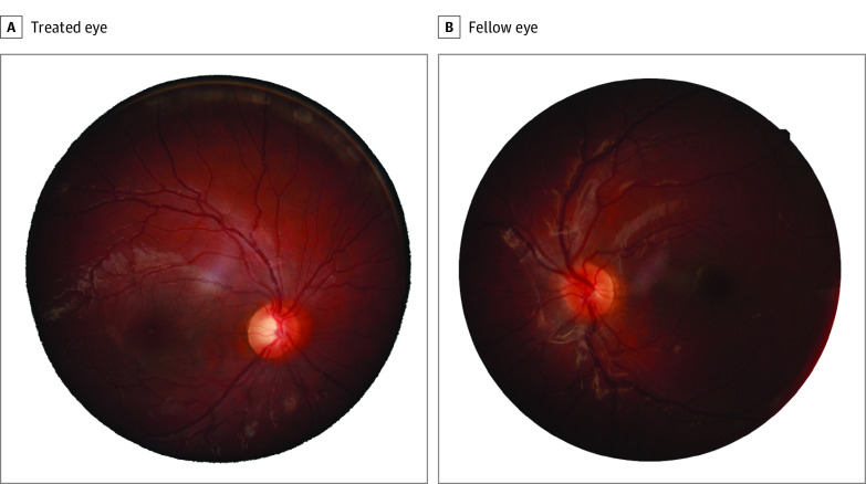
Figure 4. Examples of Optic Nerve Head (ONH) Images for the Treated (A) and Fellow Eye (B) of the Same Infant Aphakia Treatment Study (IATS) Participants Whose ONH Optical Coherence Tomography Images Are Shown in eFigure 4A and B in the Supplement.
The treated eye was diagnosed as having glaucoma based both on the IATS criteria (eTable 1 in the Supplement) and ONH imaging, while the fellow eye was graded as normal (neither glaucomatous nor glaucoma suspect). The peripapillary retinal nerve fiber layer for the ONH of the treated eye (eFigure 4A in the Supplement) was 105 μm; the peripapillary RNFL for the ONH of the fellow eye (eFigure 4B in the Supplement) was 117 μm.

