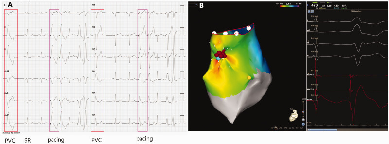
Figure 3.
Representative recordings showing the use of a notched unipolar electrogram (N-uniEGM) in the ablation of premature ventricular contractions originating from the right ventricular outflow tract (RVOT-PVC). (A) 12-lead surface electrocardiograms of sinus rhythm (SR), PVC and pacing. In this patient, the surface electrocardiogram indicated that the PVC originated from the RVOT. (B) Activation mapping of the origin of the PVC in the RVOT using the CARTO® 3 System with simultaneous recordings of surface electrocardiogram, unipolar and bipolar electrograms. The earliest ventricular activation (EVA) site was proved to be located near the anterior septum of the RVOT by 3-dimensional electroanatomic mapping. Pacing mapping morphology at this site coincided perfectly with the spontaneous PVC. An N-uniEGM was also recorded at the EVA. Radiofrequency energy delivery for 2 s at this site terminated the clinical PVC. The colour version of this figure is available at: http://imr.sagepub.com.

