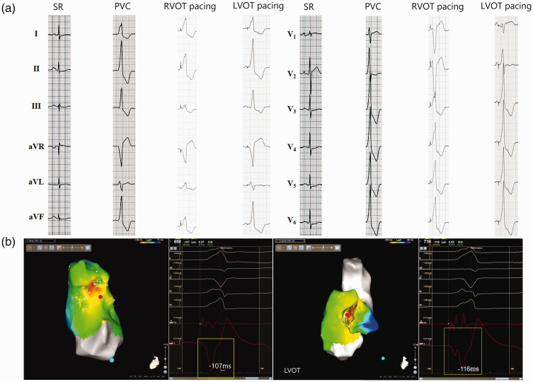
Figure 4.
Representative recordings showing the use of a notched unipolar electrogram (N-uniEGM) in the ablation of premature ventricular contractions originating from the left ventricular outflow tract (LVOT-PVC). (a) 12-lead surface electrocardiogram of sinus rhythm (SR), PVC and pacing at the right ventricular outflow tract (RVOT) and LVOT. (b) Activation mapping of the PVC target from the RVOT and LVOT using the CARTO® 3 System with simultaneous surface electrocardiogram recordings, bipolar and unipolar electrograms at the same sites and uniEGM. In this patient, the initial activation mapping at the RVOT revealed that the earliest ventricular activation site (EVA) was in the septum of the RVOT, which preceded the onset of surface QRS complex for 107 ms. Pacing mapping at this site produced a QRS morphology with a low similarity with the morphology of spontaneous PVC. The uniEGM at this site presented a QS morphology with a blunt initial part and no characteristics of N-uniEGM. Ablation at this site did not show any impact on the PVC. Activation mapping at the LVOT revealed an earlier EVA site (–116 ms) in the right coronary sinus and pacing mapping at this site produced a QRS morphology with better similarity with the spontaneous PVC. At this target site, unipolar mapping showed a characteristic N-uniEGM. PVCs disappeared after ablation at this site for 4.6 s. The colour version of this figure is available at: http://imr.sagepub.com.

