Figure 5.
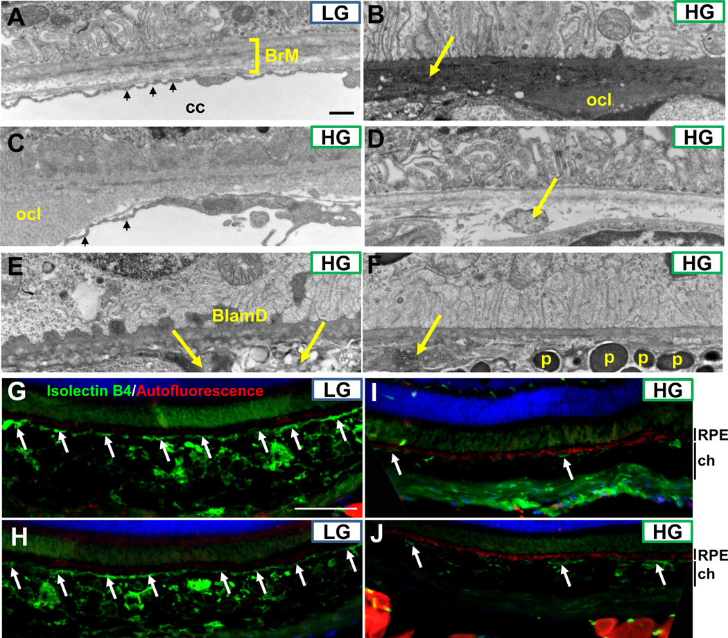
Sub-RPE deposits, Bruch’s membrane abnormalities, and loss of choriocapillaris in Nrf2-HG eyes. (A-F) Electron micrographs of the basal RPE, Bruch’s membrane, and choriocapillaris indicate the lack of sub-RPE deposits, intact penta-laminar Bruch’s membrane structure (square bracket in A), and well-fenestrated choriocapillaris (small arrows in A) in Nrf2-LG eyes (A). Nrf2-HG eyes contain multiple sub-RPE deposits, including outer collagenous layer deposits (B,C), basal laminar deposits (E), and other heterogeneous deposits/debris (arrows in B, D, E, F). The choriocapillaris was largely absent except in a few areas (small arrows, C) and choroidal tissue, including melanocytes containing pigmented granules, were localized proximal to Bruch’s membrane (F). (G-J) Immunofluorescent staining for blood vessels via IsolectinB4 staining (green) or for autofluorescence (red) indicate intact choriocapillaris (arrows) and choroidal blood vessels in Nrf2-LG eyes (G,H) that are absent or degenerated in Nrf2-HG eyes (I,J), whereas autofluorescent puncta are highly increased in Nrf2-HG RPE (red staining in I,J). Abbreviations: BlamD-basal laminar deposit, BrM-Bruch’s membrane, cc-choriocapillaris, ch-choroid, ocl-outer collagenous layer deposit, p-pigment granule within melanocyte. Scale bar in (A) is 500nm; in (G) is 100 μm.

