Figure 3.
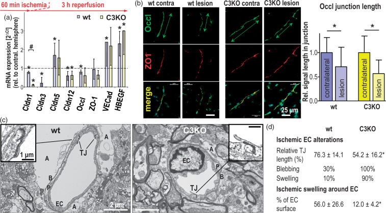
Acute ischemic injury (60 min middle cerebral artery occlusion followed by 3 h reperfusion) alters the expression of tight junction (TJ) proteins and their junctional localization in cerebral capillary endothelial cells (EC), resulting in more EC damage but less perivascular lesions in claudin-3 knockout (C3KO) than in wild type (wt) mice. (a) mRNA normalized to 28S ribosomal RNA (28SRNA) in postischemic brain capillaries excised by laser capture microdissection. Mean ± SD, n = 4; *p < 0.05 compared to the respective value in the contralateral hemisphere (dashed line) and #p < 0.05 vs. wt, using Mann–Whitney test. Cldn: claudin; Occl: occludin; ZO-1: Zonula occludens protein 1; VECad: vascular endothelial cadherin; HBEGF: heparin-binding epidermal growth factor-like growth factor (used as an indicator of ischemic injury45). (b) Immunohistochemistry of Occl showed decreased localization in cell contacts (reduced length of Occl-positive junctions measured along the vascular axis); ZO-1 served as cell contact marker. Mean ± SD, n = 41–61; *p < 0.05. (c) Transmission electron microscopy of the postischemic neurovascular unit exhibited reduced TJ length, stronger EC swelling and less swollen astrocytic endfeet in the lesional region of the C3KO brain compared to the respective wt brain; A: astrocytic endfeet; B: basement membrane; P: pericyte. (d) Quantification of the ultrastructural alterations in the vascular part of the neurovascular unit due to acute ischemic injury. The swelling around capillary endothelial cells was determined as the proportion of swollen astrocyte endfeet surrounding the abluminal part of the capillaries in electron microscopic images. Mean ± SD, n ≥ 10; *, p < 0.05.

