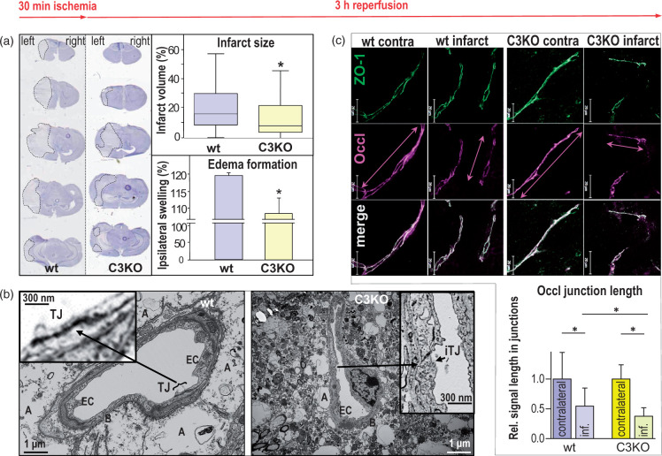
Figure 5.
Forty-eight hours after 30 min transient middle cerebral artery occlusion (tMCAO), reduction in infarction, swelling, tight junction density and length of junctional occludin (Occl) is caused in brains of claudin-3 knockout mice (C3KO) compared to that from wild type (wt) brain. (a) Hematoxylin staining (left panels) showed lower infarct size (dotted lines) and ipsilateral swelling in C3KO, as confirmed quantitatively (right diagrams). Mean ± SD, n = 21; p < 0.05; Mann–Whitney test. (b) In transmission electron microscopy of cross-sectioned brain capillaries from the striatum, the tight junctions (TJs) between endothelial cell (EC) surfaces were weakened after tMCAO in the infarct area of C3KO compared to TJs in the infarct area of wt. In wt, TJs were identified in 90% of the capillaries (left micrograph), whereas TJs were detectable in 43% of the capillaries from C3KO only. In C3KO, the junctions could hardly be identified (right micrograph) due to impairment of TJs and cell membranes. The latter is accompanied by less swollen astrocytic endfeet. A: astrocytic endfeet; B: basement membrane; iTJ: intermittent tight junction. (c) In immunohistochemistry, the length of Occl-positive segments in cell junctions (Occl junction length, measured along the vascular axis) was shorter in the C3KO infarction region than in the wt infarction area (compare double arrows); cf. lower part of (c) for quantification (inf., infarct). Zonula occludens protein 1 (ZO-1) was used as a junction marker protein. Scale bars, 20 μm; mean ± SD, n = 47–60; p < 0.05; Kruskal–Wallis test with Dunn’s multiple comparison.

