Figure 1.
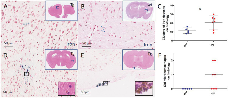
Histopathological evidence of leakage in the brains of 20-month-old APP/PS1 mice. Representative examples of areas with focal iron-positive deposits on a Prussian blue-stained section of the post-mortem brain of a 20-month-old Tg APP/PS1 mouse (a) and an age-matched WT mouse (b). A higher number of areas with clusters of focal iron-positive deposits was observed in Tg compared to WT mice (c, t-test p = 0.029). Areas with clusters of >20 focal iron-positive deposits (d, e) with evidence of hemosiderin-containing macrophages on the adjacent H&E-stained section (insets) were rare and only observed in Tg mice (f, Mann–Whitney U test p = 0.065). Lines and error bars in C indicate mean and standard deviations. Lines in F indicate median. Note that each dot in C and F represents a mouse. *p < 0.05.

