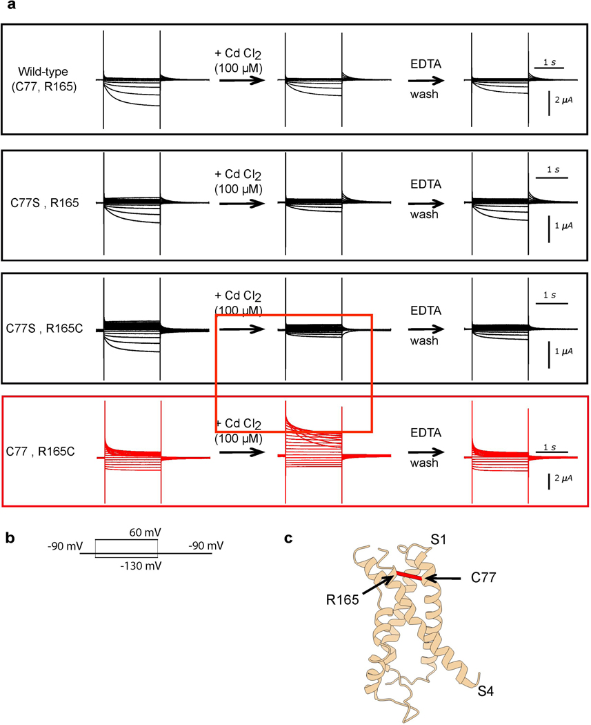
Extended Data Figure 9: A cysteine-Cd2+-cysteine bridge in the KAT1 VSD promotes channel opening.
a, Raw current traces for all four combinations of C77(S) and R165(C). Upon washing with 100 uM CdCl2, current increases only in the C77-R165C condition (red box, middle panel), and then decreases again upon EDTA wash. Representative data are shown from the same oocyte, and each experiment was repeated five independent times (five biologically independent oocytes) with similar results. b, Pulse protocol used during experiment c, Mapping of C77 (on S1) and R165 (on S4) onto the ‘up’ VSD structure of KAT1. Alpha carbons are indicated by a red line.

