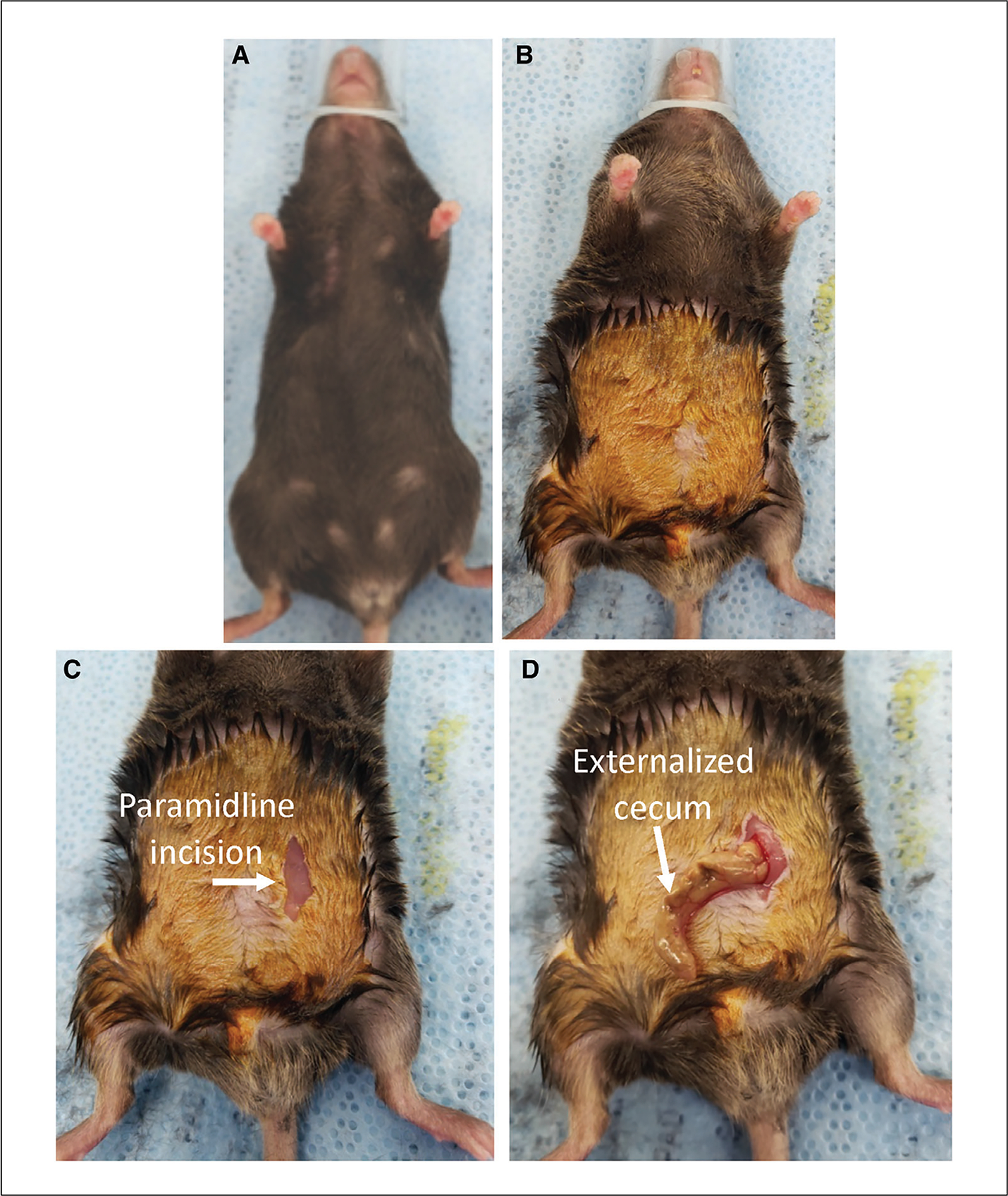
Figure 2.

Prepping the mouse and exposing the cecum. (A) The mouse is positioned on heated surgical mat and the nose cone secured. (B) The abdominal fur is shaved and 5% povidone-iodine antiseptic is applied. (C) A small (~1 cm) paramidline incision is made through the skin. (D) The cecum is externalized.
