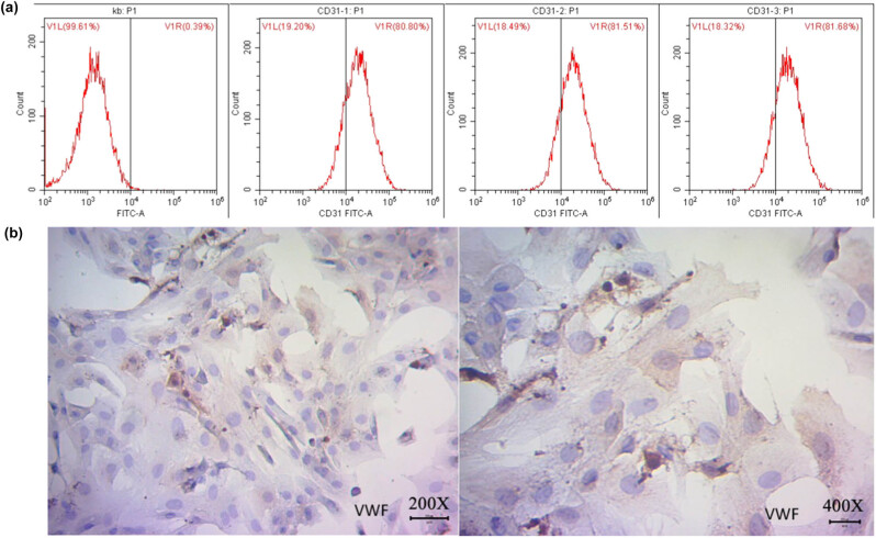
Figure 1.
Isolation and identification of RMECs from RRVECs. RRVECs were isolated and cultured from the retina of a 7-day-old neonatal SD rat, and RMECs were verified after three passages and flow cytometry. (a) Verification of RMECs using flow cytometry with CD31 antibody. Kb - the blank control without antibody; P1 - the cell content after removal of cell debris; V1L - the content of the negative cells which were not labeled by CD31; V1R - the content of the CD31 positive cells, RMECs > 80% among RRVECs. (b) Immunological verification of RMECs with a polyclonal antibody against the VWF.

