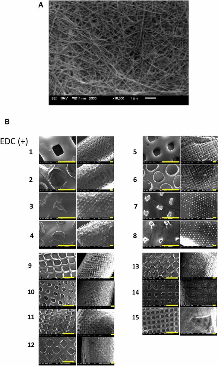
Figure 3.
(A) Scanning microscopic image of the collagen matrix crosslinked with 1% EDC. The diameter of 1% tilapia scale collagen fibrils after crosslinking was ranged from 40 to 120 μm (n = 5) although it was 30 to 100 μm before crosslinking (compared with the Fig. 3(a) in our previous study15. (Magnification 10,000 × ; Scale bar = 1 μm). (B) Scanning microscopic images of the collagen scaffolds with 15 different micropatterns’ design shown in Fig. 1. (Magnification: Left panels 1–8 300 × , 9–15 200 × , Right panels 50 ×) (Scale bar = 200 μm).

