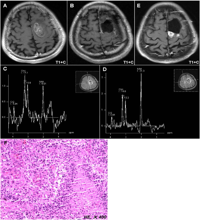
Figure 1.
Representative case of this study. Preoperative MR images showed a lesion located in the left anterior central gyrus and supplementary motor area (A). Postoperative MR images displayed that the tumor has been totally removed (B), while the postoperative peritumoral zone showed a higher ratio of Cho/NAA (>1.31) than that in the contralateral region (C,D). Twelve months after operation, the MR images showed a new enhanced lesion in the peritumoral zone (E), which was further confirmed as tumor recurrence by biopsy (F).

