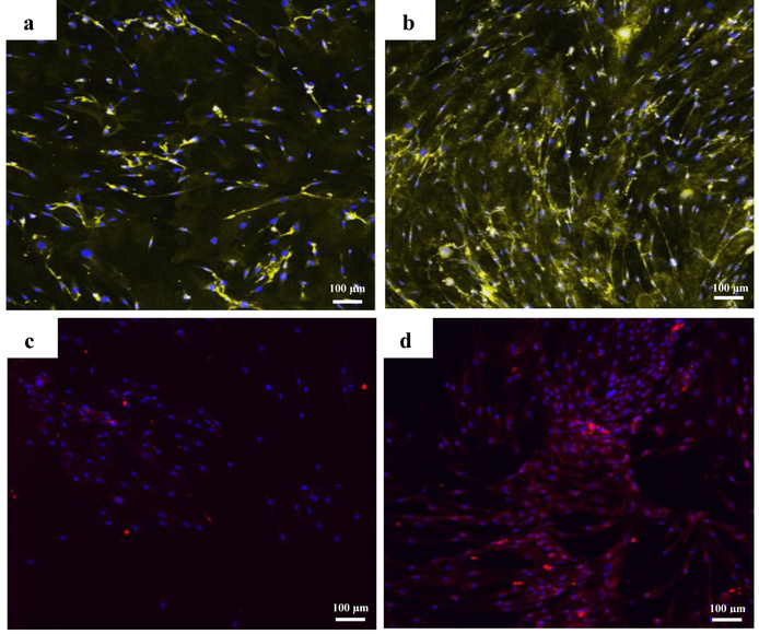
Fig. 5.
Immunofluorescence staining against vWF (yellow) on a) non differentiated cells and, b) endothelial differentiated cells and against VE Cadherin (red) on c) non differentiated cells and, d) endothelial differentiated cells after 14 days in 2D culture. The nuclei were counterstained with DAPI.

