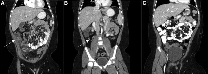
FIGURE 1.

CT scan with oral and intravenous contrast of the abdomen and pelvis. A and B, CT scan of the abdomen and pelvis on presentation showing extensive phlegmonous changes in the right lower quadrant of the abdomen around the ileocecal junction (white arrowhead) with intact appendiceal wall (dashed arrowhead). C, CT scan of the abdomen and pelvis demonstrating complete resolution of phlegmonous ileocolitis after treatment. CT = computed tomography.
