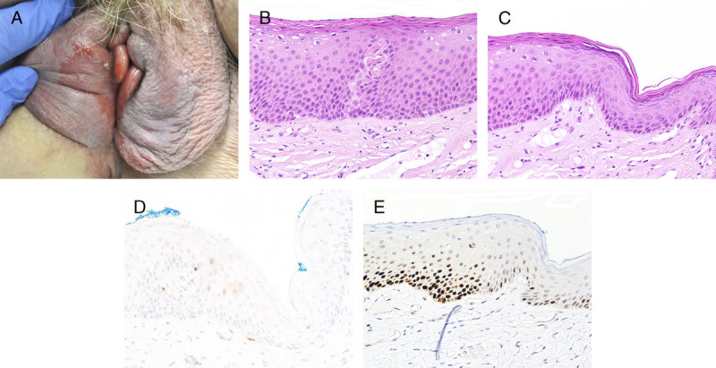
FIGURE 3.

A, Differentiated vulvar intraepithelial neoplasia—large glazed red patch over bilateral labia minora and right interlabial fold. B, Subtle keratinizing dVIN with PK, flat acanthosis, and mildly hyperchromatic enlarged nuclei extending halfway up the epithelium, H&E ×200. C, Junction between dVIN and thinner nonneoplastic epithelium, H&E ×200. D, p16 is negative, ×200. E, Basal and suprabasal overexpressed p53 in dVIN contrasts with wild-type pattern in adjacent epithelium, ×200.
