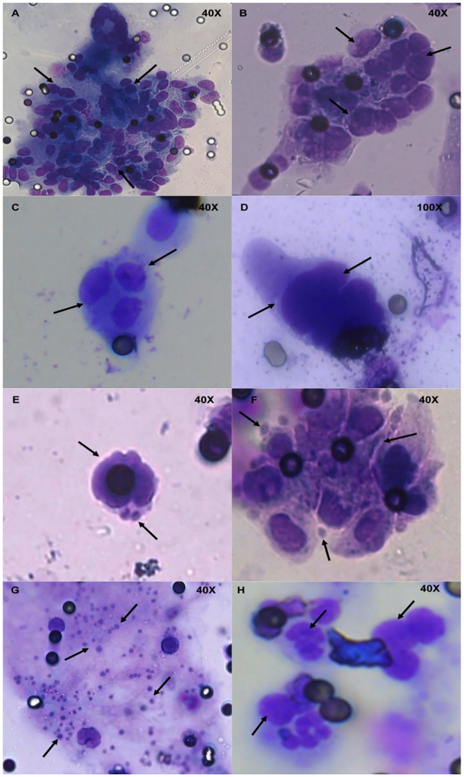
Fig 2. MGG morphological staining of representative ExPeCT filters.
A) and B) arrows depict large groups of putative CTCs (CTC clusters) and filter pore, C) arrows indicate a group of four putative CTCs, D) arrows illustrate multiple CTCs without cytoplasm attached, E) and F) arrows depict suggested platelet cloaking of CTCs, G) arrows represent dispersed single platelets on a filter and H) arrows detail inflammatory cells present in a separate plane of focus. All images were taken using a 20x or 40x objective lens, filters pores measure 7.5 μm in diameter. MGG–May Grunwald Giemsa, CTC–circulating tumour cell.

