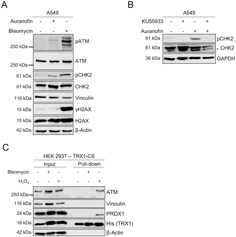
Fig 4. Auranofin induces ATM-dependent phosphorylation of CHK2.
(A) A549 cells were treated with Auranofin (10 μM) or Bleomycin (100 μM) for 6 hours or left untreated. Phosphorylation of ATM, CHK2 and H2AX was analyzed by SDS-PAGE and Western blotting. Vinculin and β-Actin served as loading controls. (B) A549 cells were treated as indicated with KU55933 (10 μM, 1 hour pre-treatment) and Auranofin (10 μM, 6 hours). Samples were subjected to SDS-PAGE and Western blotting and CHK2 phosphorylation was assessed with GAPDH as loading control. An unspecific band is marked with an asterisk (*). (C) HEK 293T cells expressing TRX1-CS were subjected to Bleomycin (100 μM, 4 hours) or H2O2 (10 mM, 15 minutes) or left untreated. Lysates were prepared in the presence of NEM and TRX1 and proteins were enriched using streptavidin-coated beads. Bound proteins were analyzed by SDS-PAGE and Western blotting. Blots were probed for ATM, PRDX1 and His-tagged TRX1. Vinculin and β-Actin served as loading controls. (A, B, C) Representative blots of two independent experiments are shown.

