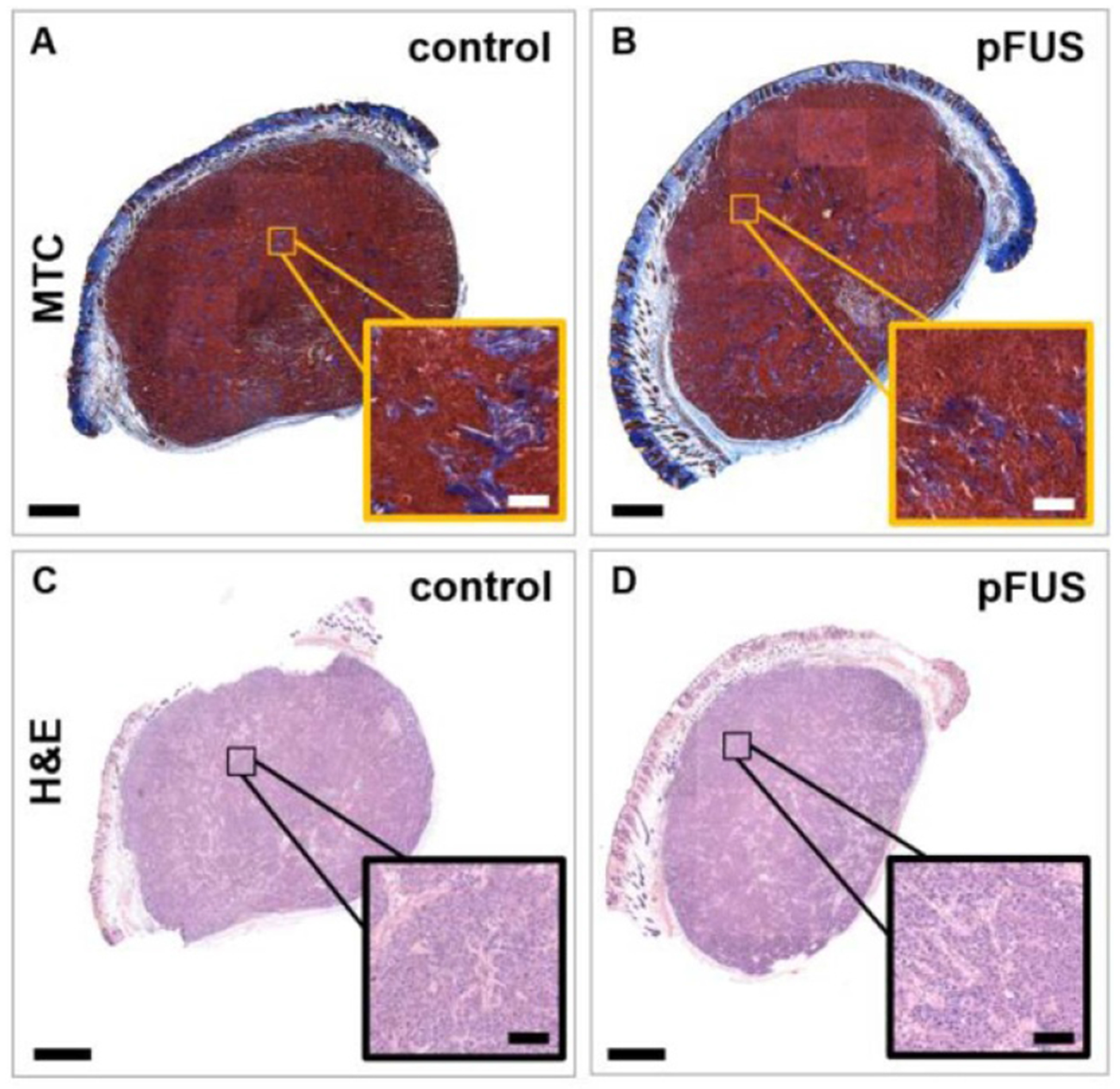
Figure 6.

Effects of pFUS treatment on the histomorphology of the tumors and fibrillar collagen network. Representative (A), (B) MTC and (C), (D) H&E-stained sections of (A), (C) control and (B), (D) pFUS-treated tumors (n = 3). Insets show zoomed-in regions. No major size, structural or histomorphological differences were observed from the stained sections based on large collagen fibers or the spatial distribution of intact cells, indicating no gross tumor damage. Bars = 1 mm and 100 μm (inset).
